Digestive System
Points to study
1)
what is digestive system
2)
part of digestive system
3)
digestive system organs and
function
4)
how does the digestive system work
5)
digestive system function
6)
digestive system process
7)
diagram of digestive system
8)
anatomy and physiology of digestive
system
9)
anatomy of organs
10)
function of organs
The
digestive system performs the following functions in our body.
1. Converts
the various food stuffs by chemical action into simpler forms which can be
easily absorbed into blood. The absorbed material is utilized by the various
tissues of the body according to their requirements.
2. This
is the pathway for the entry of vitamins, fluids and minerals into our body.
3. The
digestive system is also responsible for the excretion of residue and waste
products.
The
digestive tract begins at the mouth and ends at the rectum. There are certain
accessory organs of digestion and they are the salivary grands, liver and
pancreas. Digestive process in the body takes place in four stages:
1) Ingestion,
mastication and swallowing
2) Digestion
proper
3) Absorption
4) Excretion
of the left out and waste products.
a)
Ingestion
or taking in of food and mastication are performed by the mouth and teeth. This
is aided by the tongue. The pharynx and Oesophagus are concerned with
swallowing.
b)
Digestion
proper although begins in the mouth, is mainly carried out in the stomach and
upper part of the small intestine.
c)
The
absorption of the various food stuffs takes place mainly in the small intestine
but it can also take place from other parts of the digestive tract depending on
the nature and state of the substances concerned.
d)
In
the large intestine absorption of water and transit of the residue for
excretion in the form of faeces takes place.
ANATOMY OF DIGESTIVE ORGANS
It
consists of the lips, mouth, pharynx, oesophagus, stomach, duodenum, jejunum,
ileum (small intestine), caecum, appendix, colon and rectum. Out of this the
lips, mouth, pharynx and oesophagus are concerned with the ingestion of food
while the stomach, small and large intestine are concerned with digestion,
absorption and excretion of food stuffs.
The
process of digestion is aided by certain accessory organs of digestion namely
the salivary glands, liver and pancreas, mainly by way of their secretions. The
splitting up of the food into simpler forms involves a number of chemical
processes which are performed by means of substances secreted by the glands of
the alimentary tract and these are An enzyme is a substance which is secreted
by the called enzymes.
glands
of the digestive tract and has the ability of acting on a food stuff to convert
it into a simple form suitable for absorption. Each enzyme, however, has a
specific action i.e. it acts on one particular type of food stuff.
The
first part of the digestive system to deal with the food stuffs is the mouth.
The
digestive process which begins here is aided by the salivary secretion from the
salivary glands. Mouth is the upper expanded portion of the alimentary canal.
The interior of the mouth is lined by mucous membrane. At the floor of the
mouth is the tongue.
Tongue -:
is a muscular organ covered on its free
surface by a special type of mucous surface. The mucous membrane of the mouth
is smooth except that of the tongue. The mucous membrane of the tongue is rough
and this is due to the numerous minute elevations called papillae.
Microscopic
examination of the mucous membrane from the sides of the tongue shows that it
contains a number of specialized collections of epithelial cells called taste
buds. The hind most part of the tongue is attached the hyoid bone. There are
three types of papillae found on the tongue
1.
Eight or nine large papillae arranged in a V-shaped manner the base of the tongue.
These are called the circumvallate papillae and contain numerous taste buds.
2.
Fungiform or flattened papillae.
3.
Filiform papillae which are small pointed elevations.
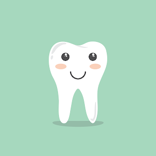
Teeth -:
During the lifetime of an individual two sets
of teeth make their appearance. The deciduous or temporary (milk) teeth and the
permanent teeth. There are 32 permanent teeth 16 above and 16 below. Out of 16
teeth, four are incisors (2 on either side), 2 canines (I on either side),
premolars on either side), and 6 molars (3 on either side).
 |
| teeth,digestive-system |
Functions of teeth -:
The
ingested food undergoes mastication and teeth is essential for this. Chewing or
mastication is the process biting and grinding of food between the upper and
lower teeth. This breaks up large food particles and mixes the food with
salvia. Mastication is assisted by tongue and cheek. The muscles of mastication
include the temporalis, masseter and the medial and lateral pterygoids.
Salivary Glands
and Salivary Secretion
-:
The
three pairs of salivary glands are:
i) Parotid
ii) Submandibular
or submaxillary
iii) Sublingual
The
salivary glands are racemose (looks like a bunch of glands). Each gland
consists of a large number of acini. Each acinus is lined by pyramidal shaped
epithelial cells. The secretion of these glands, saliva enters the mouth by
means of the ducts of the glands. Fine ductless which unite with similar ductless
to form a bigger ductless. Similar bigger ductless unite and ultimately the
main duct of the gland is formed.
All these glands receive nerve supply from
sympathetic and parasympathetic divisions of the autonomic nervous system.
Parasympathetic innervation is more important than the other. Salivary glands contain
either mucous or serous cells or both. Mucous cells contain mucinogen granules
and serous cells zymogen granules.
Functions of
Saliva -:
1)
Its
main function is to help digestion and to provide a protective secretion in the
mouthy which keeps the mucous membranes moist and these aids in oral hygiene.
2)
Lubricating
the food in preparation for swallowing.
3)
Saliva
acts as solvent for the molecules which stimulate the taste buds and has
cleansing action.
4)
It
helps in maintaining the oral pH and also helps in regulation of water balance.
Stomach -:
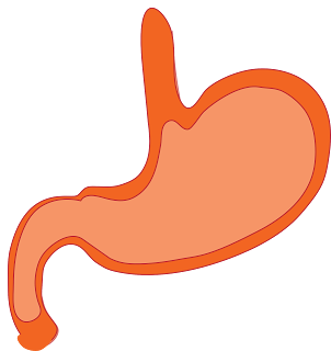
Stomach
acts as a receptacle for food. Here the food is stored where it is mixed with
acid, mucous and pepsin and released at a controlled and steady rate into the
duodenum. It is the most dialated portion of the digestive tract and lies under
the diaphragm.
 |
| stomach,digestive-system |
The stomach has three parts:
a. The
fundus which is an upper portion ballooning towards the left;
b.The
body which is the central portion; and
c. The
pyloric portion (antrum), a relatively constricted portion at the terminal part
with pylorous, just before the entrance into the duodenum. The structure of
stomach
Its
wall has four coats namely:
· Serous
· Muscular
· Submucous
· Mucous
The
muscular layer can be further differentiated into three layers. From outside to
inwards, these are:
1. Longitudinal fibers
2.
Circular fibers
3.
Oblique fibers
The
peritoneum forms the outer covering of the stomach. The musculature is heavier
in the pyloric portion than in the rest of the stomach. The circular muscle
layer is thickened in the pyloric portion to form the pyloric sphincter.
When the stomach is empty the mucous membrane
is thrown into prominent folds called rugae. These become flattened when the
stomach is full. The epithelium of the stomach is simple columnar. Gastric
glands, densely packed numbering about 35 million open at the surface by way of
gastric pits. The glands are branched tubular and penetrate the lamina propria
all the way to the muscularis mucosae.
Functions of Stomach -:
1. Storage of food -:
When food enters stomach, it
relaxes by a reflex process of receptive relaxation. This relaxation is aided
by the movement of the pharynx and oesophagus. The food is stored here until it
is emptied into the duodenum.
2. Gastric secretion -:
This is the secretion of the
gastric glands present in the stomach, The gastric secretion contains enzymes
(see Table 10.1) and mucus. Two other important components of the gastric juice
are hydrochloric acid and pepsinogen.
3.Hydrochloric acid secretion -:
Hydrochloric acid is secreted by
parietal cells of the gastric glands. It is concentrated enough to cause tissue
damage, but in normal individuals the mucosa does not get damaged because of
the surface membrane of the mucosal cells.
4.Pepsinogen secretion -:
Chief cells of the gastric
glands secrete pep- sinogens which are the precursors of the pepsins in the
gastric juice.
5. Mixing of food with acid mucus and pepsin -:
Peristaltic contractions are responsible for the mixing of food with acid,
mucus and pepsins.
6.Gastric emptying -:
Peristaltic contractions in the
stomach, in addition to mixing of food is also responsible for the entry of
food into the duodenum at a controlled rate, Peristaltic wave which may be upto
3 min are marked most in the distal half of the stomach. Pyloric sphincter has
only a limited control in gastric emptying. of duodenum function as a unit and
contract in a sequential manner. This allows the gastric contents to enter the
duodenum in bits.
The antrum, pylorus and upper part Gastric
secretion and motility
are regulated by neural and hormonal mechanisms.
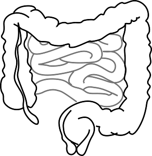
Small Intestine -:
This
extends from the distal end of the pyloric sphincter to the cecum which is the
first portion of the large intestine.
It
is about 18 feet in length and is divided into three parts namely:-
1.
Duodenum
2.
Jejunum
3.
Ileum.
 |
| small-intestine,digestive-system |
Duodenum -:
It is the shortest, widest and
most fixed portion of the small intestine. It is about 22 cm. in length.
Jejunum -:
This is about two fifths of the
small intestine.
Ileum -:
This constitutes three fifths
of the small intestine.
Both
small and large intestine is anchored to the abdominal wall by folds of
peritoneum called mesenteries.
Structure of Small Intestine -:
The four
layers typical of the gastro intestinal tract is present in the intestine also.
However, there are certain specialized features particularly in the mucosa. The
small intestine is concerned with absorption and hence this modification helps
to increase the surface area. The following are seen:
1. Circular folds (Plicae circulares) -:
These are large permanent, transverse folds
seen in the entire thickness of the mucosa containing a core of sub mucosa.
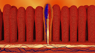
2. Villi -:
 |
| villi,digestive-system,villus |
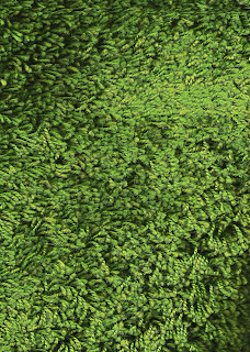
Villi are finger like projections
of the mucosa into i lumen containing blood vessels and a centrally located
lymphatic vessel (central lacteal).
3. Microvilli -:
 |
| digestive-system,villi,microvilli |
There are many cylindrical
processes on the free edges of the epithelial cells which give a striated or
brush border Crypts of Lieberkuhn are the tubular glands found in the mucosa
between the villi.
In the
duodenum there are small coiled glands which secrete mucous high in bicarbonate
content. These are called Brunner's glands. Simple columnar epithelial cells
are found lining the small intestine. Amidst the cells of the epithelium are
found goblet cells which secret mucus.
Functions
of Small Intestine
-:
1) Secretion
of the small intestine contains many enzymes which help in digestion of various
food stuffs. Pancreatic secretion is also poured into the small intestine along
with bile secretion) this is also helpful in digestion.
2) Most
of the absorption takes place at the small intestinal mucosa)
3) Various
types of movements of the small intestine like peristalsis. segmenting
contraction help in the mixing and churning of food materials before propelling
it towards large intestine.
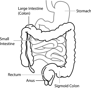
Large Intestine -:
Large intestine is approximately five feet
long and extends from the end of ileum to the anus and is divisible into:
i. Cecum;
ii. Colon;
iii. Rectum; and
iv. Anal canal
 |
| large-intestine,intestine,digestive-system |
It is different
from the small intestine in the following ways:
1. It is wider
than the small intestine.
2. Its mucosal
surface has no villi.
3. In the cecum,
colon and upper part of the rectum the glands present are of greater depth and
more closely packed. This contains more of goblet cells.
Cecum -:
This is the first
portion of the large intestine. Attached to the base of cecum is a slender
tube; the verniform (worm like) process or appendix. lleocaecal valve is
present at the junction of ileum and cecum.
Appendicitis or inflammation of the appendix
occurs in both children and adults.
Ascending colon -:
It extends
upward from the cecum.
Transverse colon -:
This
overlies the coils of the small intestine and Ascending colon crosses the
abdominal cavity from right to left below the stomach.
Descending colon -:
This
begins near the spleen passing downward on the left side of the abdomen to the
iliac crest to become the sigmoid pelvic colon. This is so called because of
its S-shaped course within the pelvic cavity.
Rectum -:
The pelvic
colon passes into the rectum and lies on the anterior surface of the sacrum and
coccyx. It terminates in the narrow anal canal which opens to the exterior at
the anus.
Structure of the Large Intestine -:
Like the other
parts of the gastro intestinal tract including small intestine, large intestine
also has the four coats. The muscle layer has a characteristic feature.
Longitudinal muscles do not form a continuous layer over the whole gut but are
arranged in three separate bands taenia coli. These bands being shorter than
the length of the large intestine the large intestine has a sacculated
appearance.
Functions
of the Large Intestine
-:
1.
It
secretes the intestinal secretion which lubricates the faeces and facilitates
their passage through the rectum and anus.
2.
The
bacteria present in the large intestine act on the various food materials which
are left without digestion and absorption by the intestine.
3.
Absorption
of water, glucose and salt also takes place in the large intestine.
4.
Excretion
of excess calcium, iron and drugs of the heavy metal type takes place from the
walls of the intestine,
5.
Segmentation
contraction of the large intestine helps in mixing the contents of the colon
while peristaltic contraction propel the contents towards rectum.
6.
A
type of contraction called mass action contraction in which k there is
simultaneous contraction over a large area of the intestine takes place in
descending and sigmoid portions. This is helpful in defecation.
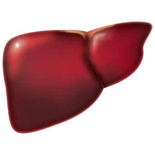
Liver -:
The
liver is the largest organ in the body and has a weight of 1275- 1550 gm. It is
situated in the upper part of the abdominal cavity immediately below the
diaphragm.
It has an irregular shape. It may be
conveniently divided into right and left lobes and has superior, inferior,
anterior and posterior surfaces. Superior surface is in contact with the under
surface of the diaphragm which separates it from the bases of the lungs.
Inferior surface is concave and is related to other abdominal viscera.
 |
| liver,digestive-system |
Anterior surface is related to the abdominal
wall in the midline. Posterior surface crosses the vertebral column in the
middle and is also related to the aorta, inferior vana cava and lower end of
the esophagus.
In
the centre of the inferior surface is the hilum or gate of the liver. This is
also called as portal fissure. This is the point of entry and exit for
(1)
Portal
vein
(2)
Hepatic
artery; and
(3)
Bile
ducts.
The
liver also has a fibrous capsule and the greater part of its surface is covered
by peritoneum.
Structure of Liver -:
A large number of lobules consisting of columns
of liver cells constitute the liver. The liver cells of a lobule are arranged
around a central vein in a radiating fashion. Each lobule has got a blood
supply from hepatic artery, and portal vein. Bile secreted by the liver cells
passes out through the bile ducts. Bile leaves the liver through the right and
left hepatic ducts. These combine together to form the common hepatic duct. The
hepatic duct is joined by the cystic duct from the gall bladder to form the
common bile duct. The common bile duct joins the pancreatic duct to open into
the duodenum at the duodenal papilla or ampulla of vater.
Functions of Liver -:
1)
It
produces bile. Bile mainly contains bile salts and bile pigments. Bile is
responsible for the digestion and absorption of food.
2)
It
is concerned with the storage of glycogen.
3)
Liver
is concerned with the production of plasma proteins/(albu- min and globulin)
which are essential for the body.
4)
The
breakdown of protein and the formation of urea.
5)
Storage
of Vitamin B12 and iron
6)
Heat
production.
7)
Destruction
of various toxic substances and drugs.
8)
Desaturation
of fats
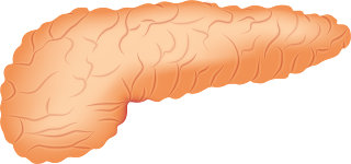
Pancreas -:
It is a large lobulated gland and resembles
salivary glands in struc- Pancreas has a head, body and tail. Pancreas has two
parts.
(1) Exocrine part
(2) Endocrine part
 |
| pencreas,digestive-system |
Exocrine part
Pancreatic juice
is secreted by the exocrine part. Pancreatic juice is alkaline and has a high
bicarbonate content. Pancreatic juice also contains a large number of enzymes
required for digestion. Pancreatic juice is secreted into the duodenum through
the pancreatic duct.
Pancreatic juice
is secreted by the exocrine part. This digestive juice contains many enzymes
for digestion. Pancreatic duct empties its secretion into the duodenum.
Pancreatic secretions is controlled by both
neural and hormonal factors. Secretin and cholecystokinin-pancreozymin
(CCK-Pz or CCK) are the two hormones controlling the secretion. Vagal
stimulation also causes secretion of pancreatic juice.
Endocrine
part -:
The Islets of Langerhans, a collection of
ovoid cells scattered throughout the pancreatic tissue (more cells in the tail
region) forms the endocrine part of the pancreas. A or α
cells secrete glucagon and B or B cells secrete insulin. The known effect of
insulin is its hypoglycemic action. Insulin facilitates the entry of glucose
into the cells except in brain. Glucagon on the other hand causes break down of
glycogen glycogenolytic action.
Physiology
of Digestion and Absorption
-:
The major
foodstuffs are carbohydrates, proteins and fats. These are broken down by the
enzymes present in the various digestive juices secreted by the different parts
of the gastrointestinal tract. Digestion of the various foodstuffs can be
described as follows:
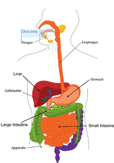
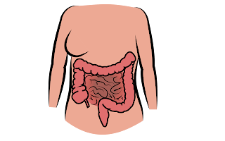 |
| digestive-organs,digestive-system |
Digestion of carbohydrates -:
The principal
carbohydrates present in food is starch composed of glucose units. Others are
sucrose, fructose and lactose. Digestion begins in the mouth with the enzymatic
action of Ptyalin, salivary amylase. The enzymatic action of ptyalin continues
in the stomach. All sugars are converted into simple monosaccharides by various
enzymes (sucrase, maltase, lactase). In the small intestine en- cymes like
pancreatic amylase, maltase, sucrase and lactase act up on the Starch and
disaccharides respectively.
Digestion of proteins -:
The digestion of
protein is initiated in the Momach by the action of pepsin of the gastric juice
and proteins ure verted into peptones and polypeptides. Trypsin and
chymotrypsin of the pancreatic juice also acts upon the proteins in the
intestine and convert them into peptides and peptidases, split peptides into
amino acids.
Digestion
of fat -:
Pancreatic
lipase splits fat into monoglycerides, fatty acids and glycerol. Intestinal
lipase also splits the fats.
Absorption
-:
The absorption of the digested food material
occurs almost exclusively in the small intestine. However, some glucose, alco-
hol and water are absorbed in the stomach. Significant amounts of water and
salts are absorbed from large intestine. The following Table shows the site of
absorption and the substances absorbed.
Mouth
normally no absorption but a few
drugs may be absorbed into the blood through the mucous membrane in case it is
allowed to dissolve under the tongue e.g. glycerol nitrate, isoprenaline.
Stomach
Water;
glucose; alcohol; and some drugs may also be absorbed.
Small Intestine
Water;
amino acids; simple sugars; short chain fatty acids; glycerol, vitamins and
minerals; calcium
Large Intestine
Products of lipid digestion.


Grrat
ReplyDeleteSuper
ReplyDelete